
Science Illustration Services
I create science illustrations for: journal covers, procedural explainers, visual abstracts, research figures and more.
As a long-time science visualizer at Columbia University and a member of the Guild of Natural Science Illustrators, I collaborate with researchers, clinicians, and institutions—including CUNY, UCLA, Boston Medical, and NY Medical College—to ensure each image is both scientifically accurate and visually compelling.
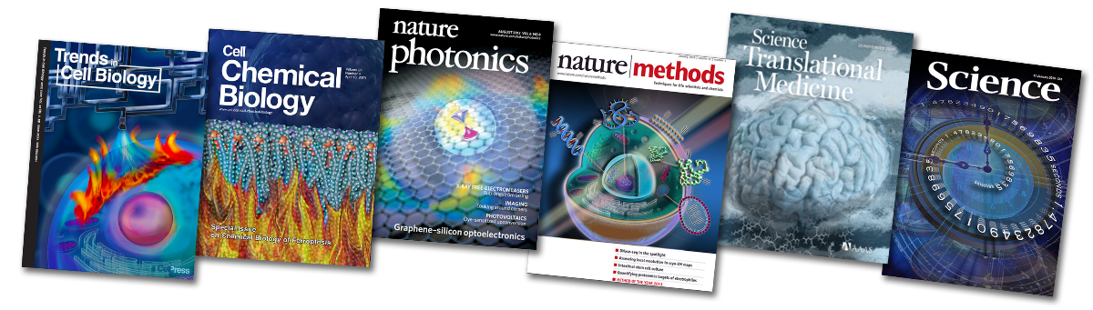
Scientific journal cover illustrations for publications including Trends in Cell Biology, Cell Chemical Biology, Nature Photonics, Nature Methods, Science Translational Medicine, and Science. Work focuses on visualizing complex biological, chemical, and physical processes for academic research.

Step-by-step surgical illustration depicting nephrectomy. Designed for New York Medical College medical training and procedural education.

Anatomical illustration of the human heart used to support medical education and procedural understanding for Columbia University Cardiology.

Medical illustration of targeted brain tumor resection, showing a small craniotomy and precise removal of a localized lesion. Created for clinical and research communication at Columbia University.

Medical illustration for Columbia Neurosurgery showing spinal instrumentation and minimally invasive surgical techniques.

Scientific illustration for research grant of gastrointestinal processes and microbiome interactions, highlighting bile acids, cellular pathways, and therapeutic targets.
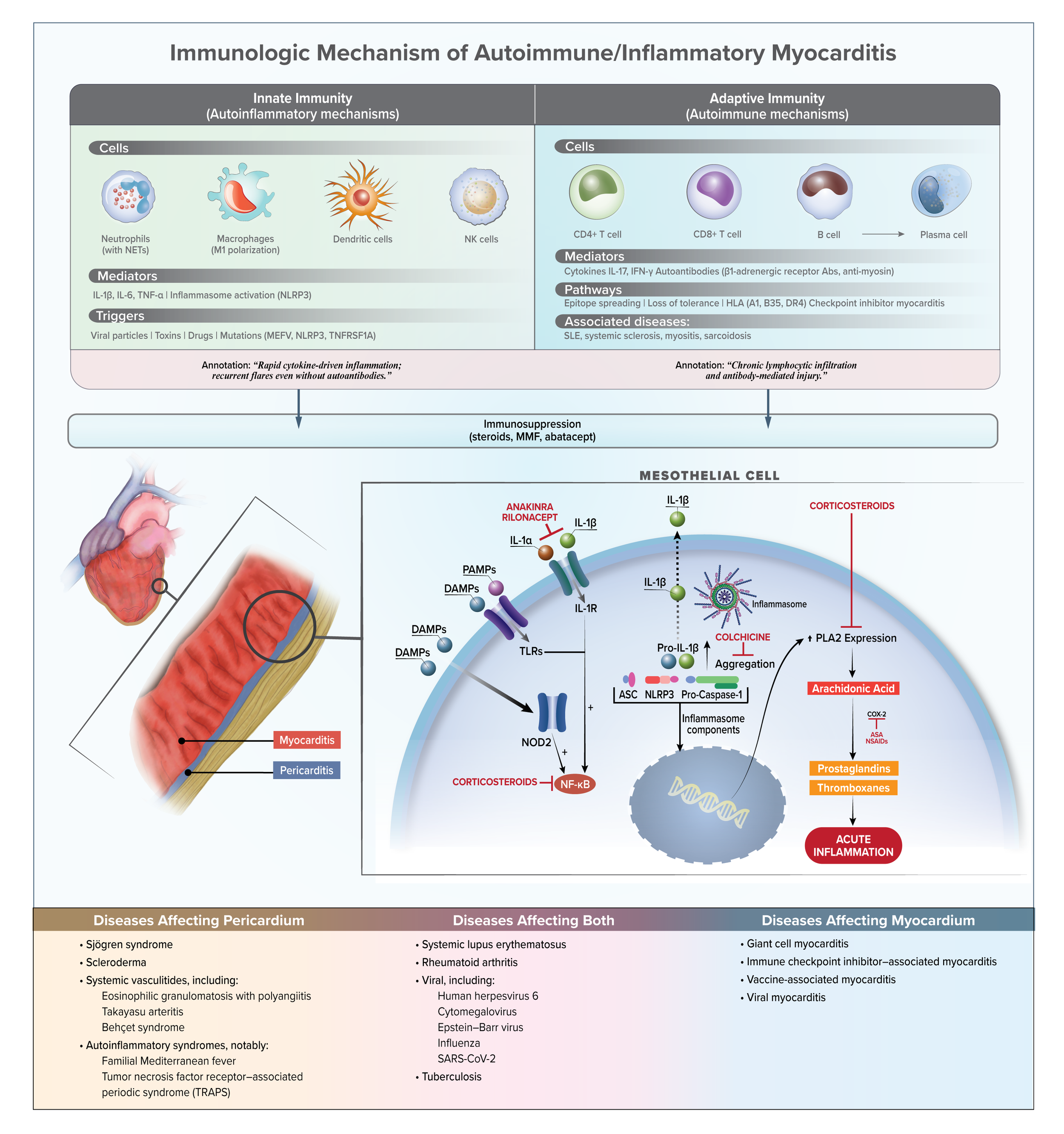
Scientific diagram of immunologic mechanisms in autoimmune and inflammatory myocarditis, illustrating innate and adaptive immune pathways, inflammatory signaling, and therapeutic targets. For research publication.

Medical illustration of non-invasive and invasive respiratory support systems, including CPAP, BiPAP, nasal cannula, and mechanical ventilation.
Designed for clinical education and patient communication Columbia University.